Tuesday, December 6, 2011

Unstable Pubic Ramus and Y-Shaped Sacral Fractures

A 72 years old female with a history of inflammatory bowel disease was injured while on vacation with her family. She fell down 4-5 steps onto a grassy lawn and noted immediate anterior and posterior pelvic pain. She was taken to a local emergency room where physical examination and plain pelvic radiographs revealed no abnormalities.

She was unable to ambulate without significant assistance and returned home. One week after injury, she consulted her primary physician and complained of worsening pelvic pain. She had no bowel or bladder symptoms.

She was referred to our clinic on that same day. On physical exam, she had pelvic mechanical instability. She had diminished strength of the hip area and lower extremity muscles bilaterally due to pain.

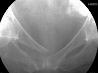
Her detailed neurological examination was otherwise normal. Pelvic plain radiographs and a computed tomography scan identified a right-sided displaced pubic ramus fracture and a displaced Y-shaped sacral fracture. On the anteroposterior image, she had a paradoxical inlet appearance of the sacrum.

The sagittal sacral CT scan images best demonstrated the displacement of the upper sacrum through the fracture’s transverse component.


She opted for surgical treatment of he unstable and displaced pelvic fractures. She underwent surgery the next day. After being anesthetized, she was positioned supine on a radiolucent table and elevated on a soft lumbosacral support. Manual compression of the pelvis under fluoroscopy identified the pubic ramus displacement due to instability.


Manipulative reduction using manual distraction at the iliac crests reduced the pubic fracture. Medullary fixation was performed percutaneously using a retrograde superior pubic ramus screw. The Y-shaped sacral fracture was stabilized percutaneously using two trans-iliac, trans-sacral iliosacral screws located safely within the upper sacral segment.


She had dramatic pain relief immediately after surgery. A licensed physical therapist monitored her rehabilitation for the next 3 months. For the initial six weeks after operation, light resistance strengthening exercises were selected along with right-sided protected weight bearing using a walker. Between weeks 7-12, progressive partial weight bearing and more vigorous strength training was performed. Four months after injury, she had returned to her normal activities without pain. Her plain pelvic radiographs demonstrated fracture union.

U, Y, and H-shaped sacral fractures are unusual and are often delayed diagnoses. The AP pelvic plain film may demonstrate a paradoxical inlet of the sacrum when the upper sacral component is displaced and kyphotic. Mid-sagittal sacral CT imaging better defines the fracture details. Percutaneous pelvic fixation is used when the fracture fragments and therefore osseus fixation pathways are adequately reduced/realigned.

Authored By: M.L. Chip Routt, Jr., M.D. 

Wednesday, November 2, 2011

Complex Monteggia Fracture Dislocation

A 25 year old male was involved in a severe motor vehicle collision in which his car collided with a transport truck.  He sustained an isolated open fracture dislocation of his dominant elbow.  On physical examination he had multiple small open wounds over the posterior aspect of his elbow and forearm.  He had a normal neurologic and vascular examination.

Radiographs were performed which demonstrated an extremely comminuted fracture dislocation of his elbow.  He had complete disruption of his proximal radioulnar joint with anterior dislocation of the radial head representing a Type I Monteggia injury variant.


The patient was brought emergently to the operating room for irrigation and debridement of his open wounds as well as open reduction and internal fixation of his fracture. He had massive disruption of the deep soft tissues and musculature around the elbow.


The joint was meticulously reduced and repaired and the ulnar shaft component was held with clamps. However, the radial head was not reduced indicating that length had not been completely restored to the comminuted ulnar shaft. Length was then added and held with a minifragment plate on the shaft of the ulna and the radial head was then reduced. A plate spanning the joint and ulnar shaft components was then placed to hold the reduction. The plate was a 3.5 mm LCDC thickness plate.


The elbow was then brought through a full range of motion and the radial head remained reduced and stable throughout the full arc of flexion and extension in full pronation and supination.

Postoperatively, the patient was started on a rehabilitation program of elbow range of motion exercises 48 hours after surgery. He was able to regain excellent motion and return to his full time manual job within six months of injury. Final radiographs demonstrated a healed ulna with a concentrically reduced joint.


Type I (anterior) Monteggia injuries are fracture dislocations of the elbow in which the ulna is fractured and the proximal radioulnar joint is disrupted. The radial head dislocates anteriorly in this type of injury. In adults, these injuries typically result from high energy mechanisms and can have a high incidence of nerve or vascular injury. The key to restoring elbow stability is anatomic reduction and length restoration of the ulnar shaft.

Most cases do not involve such an extensive fracture of the joint as was seen in this patient. However, like other Monteggia injuries, anatomic ulnar alignment was the key to restoration of elbow joint stability in this case. This patient had an excellent result which was only possible by accurately restoring his anatomy and placing sufficient sized implants to ensure a rigid and stable construct that allowed for early range of motion and elbow rehabilitation.

Authored By: Daphne Beingessner, M.D. 

Tuesday, October 4, 2011

Anterior Column

A 54 years old male fell approximately four feet from a ladder onto the ground. He complained of left hip pain and was unable to stand. On presentation to the emergency department, he was awake and alert, and only complained of severe left hip pain.

He was hemodynamically stable and his physical examination confirmed left hip pain with any attempted passive movement. There was no obvious limb deformity or abnormal other findings. The injured lower extremity’s neurological and vascular examinations were within normal limits, yet the muscle power evaluation was limited due to pain.


Pelvic radiographs and a computed tomography scan demonstrated a displaced left anterior column acetabular fracture. The axial CT images demonstrated the details including the dome involvement. Three-dimensional surface rendered images further defined the osseus anatomy of the fracture.

The fracture line extended from the posterior aspect of the iliac crest, through the iliac fossa, along the pelvic brim, and divided both the acetabular dome and anterior acetabular wall regions in half. Skeletal traction was used to protect the femoral head and acetabular fracture surfaces, relax muscle spasm, provide comfort, and alert the ancillary staff to the injury before surgery.


One day after injury, the patient underwent open reduction and internal fixation of the fracture using an ilioinguinal surgical exposure. The fracture surfaces were cleansed and then the fragments manipulated and clamped. The clamp was applied onto the pelvic brim and quadrilateral surface using the Stoppa interval of the ilioinguinal exposure.

A malleable plate and interfragmentary lag screws were used to definitively stabilize the fracture. The plate was secured initially onto the stable iliac bone and located just lateral to the sacroiliac joint. Then lag screws were inserted through the plate and between the tables of the iliac crest bone.

The antegrade medullary superior pubic ramus lag screw was inserted percutaneously safely within the osseus fixation pathway using fluoroscopic biplanar imaging. On the postoperative anteroposterior pelvic image, the surgical staples indicate the ilioinguinal skin incision.


On the first postoperative day, the patient began his rehabilitation program with passive range of hip motion and isometric muscle strengthening exercises. He used crutches to protect and unload the hip repair during ambulation for 6 weeks after surgery. During weeks 7-12, a routine conditioning and strengthening program was used along with progressive weight bearing.

At his follow up evaluation 3 months after surgery, he had no complaints nor limp, his hip range of motion was symmetrical with his uninjured side, and his hip and lower extremity strength had returned to normal. He returned to his regular job four months after surgery as a laborer.

Authored By: M.L. Chip Routt, Jr., M.D.

Sunday, September 11, 2011

Distal Humerus Fracture

43 year old female fell on the ice sustaining an intraarticular distal humerus fracture. This is a complex break of the end of the humerus thus involving the elbow joint. The elbow joint was in several pieces. The patient is an active, healthy female who is right hand dominant. Radiographs demonstrate a comminuted (multifragment) fracture of the distal humerus.


Standard radiographs are challenging to interpret, so additional xrays are taken with gentle traction applied to the arm. This helps delineate the fracture fragments.


A posterior approach was utilized to access the elbow. Given the severity of the fracture, including the multiple articular (joint) pieces, an olecranon osteotomy was performed to allow for visualization of the joint. The olecranon is the proximal part of the ulna. Posteriorly, it covers and contains the distal humerus, forming part of the hinge of the elbow joint. An olecranon osteotomy is a surgical procedure in which the olecranon (proximal ulna) is broken in a controlled manner and repaired at the end of the operation. With the olecranon osteotomy complete, the multiple pieces of the distal humerus fracture are fixed anatomically and secured with plates and screws.


Once the distal humerus is repaired, the final part of the operation involves fixing the olecranon osteotomy with a plate and screws.


At one year, the patient is pain free with near full range of motion. She lacks the last 5 degrees of extension and flexes to 135 degrees. She has full pronation and supination. She is back to her usual activities.


Distal humerus fractures are complex injuries which are challenging to fix and can result in significant functional limitations. When dealing with these injuries, it is important to properly identify the complexity of the fracture and adhere to good surgical principles and techniques to maximize patient outcomes.

Authored By: Lisa A. Taitsman, M.D.

Tuesday, August 2, 2011

Geriatric Hip Fracture

An active 89 year old female presented to our Emergency Department following a fall complaining of left hip pain and an inability to walk. Radiographs revealed an intertrochanteric hip fracture.


She was admitted to the hospital and evaluated by the internal medicine team. She was cleared for surgery and on the following day she was taken to the operating room for closed reduction and internal fixation with a Dynamic Hip Screw (DHS).


Following the surgery, she was allowed to put weight on her leg as she tolerated. Her immediate postoperative course was uncomplicated. Her xrays at six weeks demonstrate good alignment and evidence of early healing.


Unfortunately, just before her three month follow up appointment, she fell again and sustained a right hip fracture.


Again, the patient was evaluated and co-managed by the medical team. This fracture was treated with a closed reduction and stabilized with a medullary hip screw. We chose to use this implant due to concerns that the fracture extended distally. For fractures that extend into the subtrochanteric region, a medullary implant is preferred over the DHS. At 2 months, the following xrays were obtained.


The patient is now almost 3 years out from her second surgery and is doing well. She uses a walker to ambulate and lives in an assisted living community. When caring for geriatric patients with hip fractures it is important to provide a quality operation, but also a team approach as to optimize patient outcome.

Authored By: Lisa A. Taitsman, M.D. 

Friday, July 8, 2011

Complex Tibia Fracture

This 55 year old female sustained an injury to her right leg after a fall from a ladder.  The patient sustained an intraarticular fracture of the distal tibia combined with a non-contiguous fracture of the distal tibial shaft.  Both injuries were closed. The short oblique fracture of the tibial shaft is shown in the injury radiographs (a, b).  Further, at the ankle joint, there is a significant fracture intraarticular injury of the distal tibia which is characterized by a spiral fracture that separates a large posterior articular segment (c, d, e).  There is a small piece of cortical comminution proximally at the posterior aspect of the fracture.


(a) (b)


(c) (d) (e)

The CT scan help to further characterize the articular injury pattern. The axial images demonstrate the posterior articular segment that comprises the vast majority of the articular surface. The plane of the fracture at the joint line is largely coronally oriented with a medial cortical exit point that extends from the medial malleolus distally (a, b). The sagittal and coronal CT reformations further demonstrate the fractures and confirm the lack of significant impaction or comminution (c, d, e). These CT scans demonstrate that the medial malleolar fracture from the AP plain radiographs is actually a large articular fracture of the posterior aspect of the medial distal tibia.


(a) (b)


(c) (d) (e)

The shaft fracture, if in isolation, would certainly be optimally managed with an intramedullary nail. The displaced articular injury, if in isolation, would be optimally managed with open reduction and internal fixation. However, the leg was initially quite swollen and open reduction of the articular surface at the initial operative intervention was felt to be risky. Further, it was felt that primary stabilization of the shaft fracture with a nail would potentially block an accurate reduction of the articular injuiry. For that reason, it was elected to fix the fibular fracture and place a spanning external fixator that provided temporary stability of both the shaft fracture and the pilon injury. The fibular was plated through a posterolateral surgical exposure. The external fixator was placed from the proximal tibia (2 Schanz pins) to the foot (transcalcaneal pin with a medial pin at the cuneiforms) to span both fractures and to reasonably center the talus relative to the tibial shaft (a, b). The repeat CT scan adds very little information and was probably unnecessary. The fractures are and the orientation of the articular injury is confirmed (c, d, e).


(a) (b)


(c) (d)


(e)

The patient elevated the lower extremity for seven days to allow adequate resolution of the soft tissues at the distal tibia.  As mentioned previously, the ideal operative plan combined open reduction and internal fixation of the articular injury combined with intramedullary nailing of the tibial shaft component.  The articular injury was approached first to prioritize the joint reduction.  The patient was positioned supine.  Given the long medial cortical exit of the fracture, a posteromedial skin incision was used to allow exposure of the posteromedial proximal aspect of the fracture.  Distally, a full thickness flap was used to allow exposure of the medial distal tibia.  An incision directly over the medial face of the distal tibia was avoided to minimize potential wound healing issues.  The fracture was cleaned from proximal to distal, allow visualization of the fracture line at the joint.  Despite a thorough removal of all hematoma from the fracture and the presence of a well-corticated fracture read, the fracture could not be perfectly reduced as judged visually and radiographically.  Therefore, an anterolateral surgical exposure was performed to allow for control and clamping at the opposite fracture exit point.  This allowed for simulataneous clamp applications and improved control of the fracture.  No identifiable block to reduction was found, however, improved fracture reduction was obtained with the additional approach.  Fixation consisted of multiple independent 2.4 mm lag screws placed perpendicular to the fracture line, and additional lag screws placed through a 2.0 mm plate along the anterior distal tibia.  The lag screws and plate were placed in a location that was felt to allow for placement of an intramedullary nail for the tibial fracture (a, b, c).


(a) (b)


(c)

An attempted closed reduction of the shaft fracture was performed but regaining length was difficult. Given the proximity of the posteromedial approach which was used for the articular injury and the shaft fracture, this incision was simply extended proximally five additional centimeters to allow for reduction and clamp placement at the shaft fracture. No significant soft tissue dissection was performed (a, b). This allowed for atraumatic nail placement without vigorous manipulation of the reduced articular injury. The clamp was left in position distally to ensure that the distal fracture did not displace (c, d). A reamed nail was placed. The three distal interlocking screws (medial to lateral, anterior to posterior, anteromedial to posterolateral) were placed. An additional lag screw was then placed posterior to the nail to support the articular fracture reduction. A small lag screw was placed at the medial malleolus (e, f). Final radiographs demonstrate the reductions of the tibial shaft as well as the distal tibial articular injury (g, h).

(a) (b)


(c) (d)


(e) (f)


(g) (h)

Initially, unrestricted range of motion exercises of the ankle joint and subtalar joint was encouraged two weeks following the definitive articular reconstruction. Weight bearing was restricted until 12 weeks given the articular injury. Healing of both fractures progressed uneventfully as demonstrated in the radiographs at 6 months (a, b, c, d e).


(a) (b) (c) (d)


(e)

Criticisms and Alternatives:
Alternatively, a short intramedullary nail could have been used to treat the shaft component of the fracture, followed by delayed open reduction and internal fixation of the articular injury. This would have a required an extremely short nail that would have terminated quite close to the proximal extent of the articular fracture posterior cortical extension. However, this approach would have allowed primary stabilization of the tibial fracture, combined with fibular fixation. An ankle joint spanning external fixator would still have been required. The shaft component of the injury pattern could have been treated with a plate at the time of reduction and internal fixation of the pilon fracture. This could have been accomplished with a direct open reduction, or with a minimally invasive technique.

Authored By: Sean E. Nork, M.D.