Friday, July 8, 2011

Complex Tibia Fracture

This 55 year old female sustained an injury to her right leg after a fall from a ladder.  The patient sustained an intraarticular fracture of the distal tibia combined with a non-contiguous fracture of the distal tibial shaft.  Both injuries were closed. The short oblique fracture of the tibial shaft is shown in the injury radiographs (a, b).  Further, at the ankle joint, there is a significant fracture intraarticular injury of the distal tibia which is characterized by a spiral fracture that separates a large posterior articular segment (c, d, e).  There is a small piece of cortical comminution proximally at the posterior aspect of the fracture.


(a) (b)


(c) (d) (e)

The CT scan help to further characterize the articular injury pattern. The axial images demonstrate the posterior articular segment that comprises the vast majority of the articular surface. The plane of the fracture at the joint line is largely coronally oriented with a medial cortical exit point that extends from the medial malleolus distally (a, b). The sagittal and coronal CT reformations further demonstrate the fractures and confirm the lack of significant impaction or comminution (c, d, e). These CT scans demonstrate that the medial malleolar fracture from the AP plain radiographs is actually a large articular fracture of the posterior aspect of the medial distal tibia.


(a) (b)


(c) (d) (e)

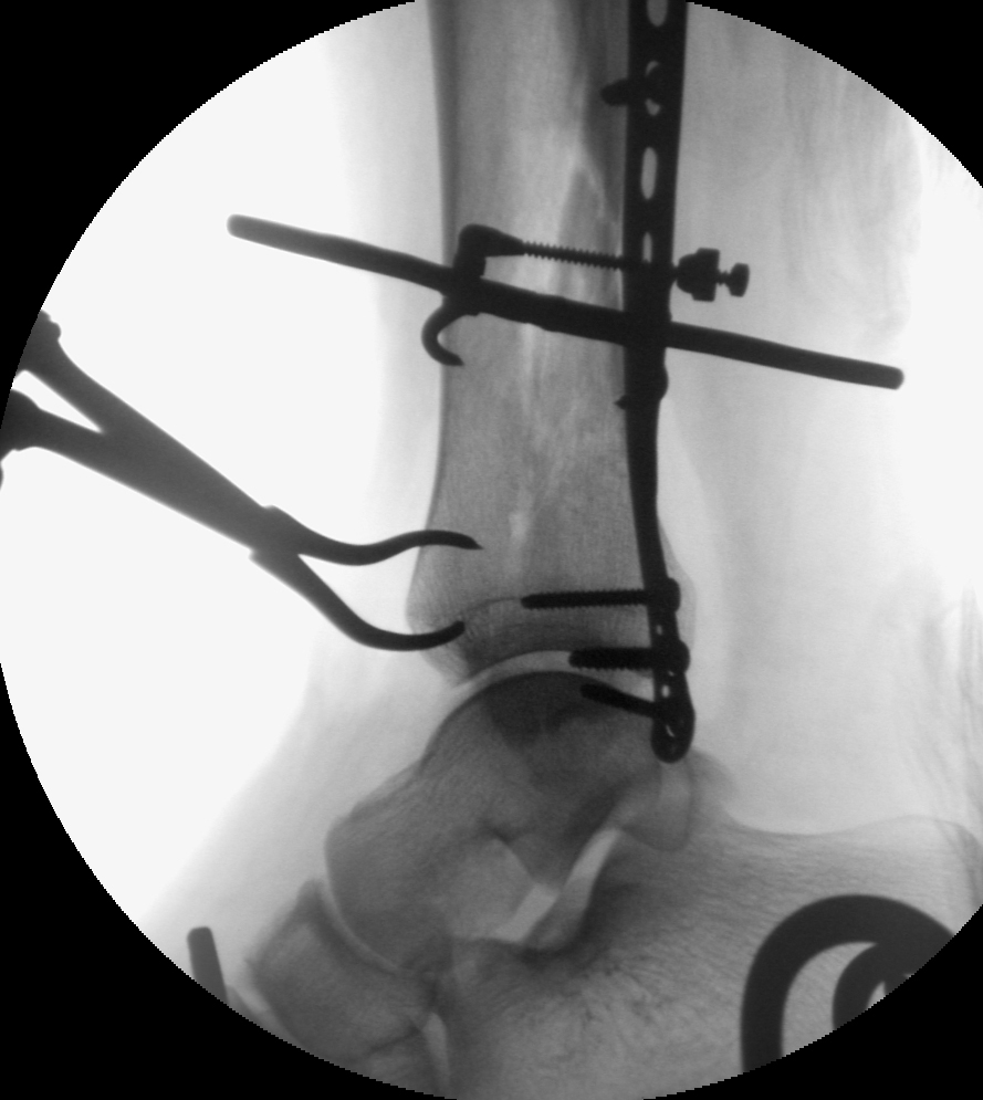
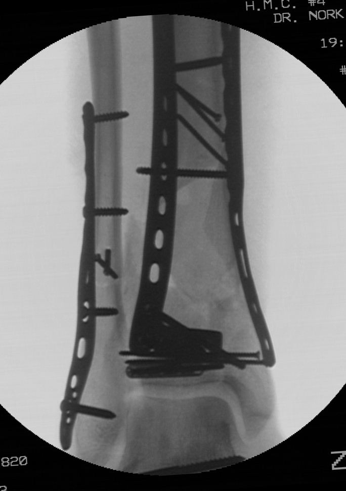
The shaft fracture, if in isolation, would certainly be optimally managed with an intramedullary nail. The displaced articular injury, if in isolation, would be optimally managed with open reduction and internal fixation. However, the leg was initially quite swollen and open reduction of the articular surface at the initial operative intervention was felt to be risky. Further, it was felt that primary stabilization of the shaft fracture with a nail would potentially block an accurate reduction of the articular injuiry. For that reason, it was elected to fix the fibular fracture and place a spanning external fixator that provided temporary stability of both the shaft fracture and the pilon injury. The fibular was plated through a posterolateral surgical exposure. The external fixator was placed from the proximal tibia (2 Schanz pins) to the foot (transcalcaneal pin with a medial pin at the cuneiforms) to span both fractures and to reasonably center the talus relative to the tibial shaft (a, b). The repeat CT scan adds very little information and was probably unnecessary. The fractures are and the orientation of the articular injury is confirmed (c, d, e).


(a) (b)


(c) (d)


(e)

The patient elevated the lower extremity for seven days to allow adequate resolution of the soft tissues at the distal tibia.  As mentioned previously, the ideal operative plan combined open reduction and internal fixation of the articular injury combined with intramedullary nailing of the tibial shaft component.  The articular injury was approached first to prioritize the joint reduction.  The patient was positioned supine.  Given the long medial cortical exit of the fracture, a posteromedial skin incision was used to allow exposure of the posteromedial proximal aspect of the fracture.  Distally, a full thickness flap was used to allow exposure of the medial distal tibia.  An incision directly over the medial face of the distal tibia was avoided to minimize potential wound healing issues.  The fracture was cleaned from proximal to distal, allow visualization of the fracture line at the joint.  Despite a thorough removal of all hematoma from the fracture and the presence of a well-corticated fracture read, the fracture could not be perfectly reduced as judged visually and radiographically.  Therefore, an anterolateral surgical exposure was performed to allow for control and clamping at the opposite fracture exit point.  This allowed for simulataneous clamp applications and improved control of the fracture.  No identifiable block to reduction was found, however, improved fracture reduction was obtained with the additional approach.  Fixation consisted of multiple independent 2.4 mm lag screws placed perpendicular to the fracture line, and additional lag screws placed through a 2.0 mm plate along the anterior distal tibia.  The lag screws and plate were placed in a location that was felt to allow for placement of an intramedullary nail for the tibial fracture (a, b, c).


(a) (b)


(c)

An attempted closed reduction of the shaft fracture was performed but regaining length was difficult. Given the proximity of the posteromedial approach which was used for the articular injury and the shaft fracture, this incision was simply extended proximally five additional centimeters to allow for reduction and clamp placement at the shaft fracture. No significant soft tissue dissection was performed (a, b). This allowed for atraumatic nail placement without vigorous manipulation of the reduced articular injury. The clamp was left in position distally to ensure that the distal fracture did not displace (c, d). A reamed nail was placed. The three distal interlocking screws (medial to lateral, anterior to posterior, anteromedial to posterolateral) were placed. An additional lag screw was then placed posterior to the nail to support the articular fracture reduction. A small lag screw was placed at the medial malleolus (e, f). Final radiographs demonstrate the reductions of the tibial shaft as well as the distal tibial articular injury (g, h).

(a) (b)


(c) (d)


(e) (f)


(g) (h)

Initially, unrestricted range of motion exercises of the ankle joint and subtalar joint was encouraged two weeks following the definitive articular reconstruction. Weight bearing was restricted until 12 weeks given the articular injury. Healing of both fractures progressed uneventfully as demonstrated in the radiographs at 6 months (a, b, c, d e).


(a) (b) (c) (d)


(e)

Criticisms and Alternatives:
Alternatively, a short intramedullary nail could have been used to treat the shaft component of the fracture, followed by delayed open reduction and internal fixation of the articular injury. This would have a required an extremely short nail that would have terminated quite close to the proximal extent of the articular fracture posterior cortical extension. However, this approach would have allowed primary stabilization of the tibial fracture, combined with fibular fixation. An ankle joint spanning external fixator would still have been required. The shaft component of the injury pattern could have been treated with a plate at the time of reduction and internal fixation of the pilon fracture. This could have been accomplished with a direct open reduction, or with a minimally invasive technique.

Authored By: Sean E. Nork, M.D.

Monday, May 30, 2011

Open pilon fracture with bone loss

This 24 year old male sustained an open right pilon fracture while skiing.  The patient was initially seen at an outside hospital and was ultimately referred approximately 16 hours following the injury.  There was a large posteromedial open wound approximately five centimeters above the ankle joint, and the proximal tibial shaft was still extruded through the open wound at presentation.  Surprisingly, the patient had intact plantar sensation and a well perfused foot.  There was obvious damage to the posterior musculature.  The injury radiographs demonstrate significant shortening and the extrusion of the tibial shaft posteriorly.


A CT scan was obtained prior to transferring the patient.  In most circumstances, the CT scan would be obtained after fibular fixation and spanning external fixation in cases where a staged approach is planned.  The injury CT scan images demonstrate the three major articular segments.  The posterior segment is large and includes the entire posterior and central portions of the articular surface, and extends to the medial shoulder of the joint.  The anterolateral articular segment extends to the medial shoulder.


The patient underwent initial irrigation and debridement, fibular stabilization, and spanning external fixation shortly after arrival to the hospital. At the same time the intercalary cortical fragment was reduced and stabilized. A posteromedial approach was used given the location of the open wound, the presence of a tibial shaft extruded through the skin, and the perceived need for debridement in this location. The incision was extended over a distance of approximately ten centimeters and was located posterior to the palpable posteromedial border of the tibia. This allowed for access to the tibial shaft, and the associated soft tissue structures. A significant portion of the flexor hallucis longus muscle was debrided and free cortical fragments were removed. The large intercalary cortical segment could be accurately reduced to the tibial shaft. This was felt to be important to decrease the ongoing pressure on the soft tissues, to allow for wound closure, and to allow for the future reduction of the articular surface. A posterolateral approach was used for fibular stabilization to allow for future anterior approaches. An external fixator was placed using a transcalcaneal pin, a medial cuneiform pin, and two tibial pins placed well proximal to the anticipated future surgical approaches. Antibiotic beads were placed into the osseus defect of the distal tibial metaphysis.


The patient was sent home for elevation and to allow for resolution of the soft tissue swelling. Twenty days after injury, the patient underwent definitive fixation of the pilon fracture. The injury pattern is amenable to fixation through either an anteromedial or anterolateral approach. Similarly, a direct anterior approach could be used. An anteromedial approach would actually allow for the best visualization of the medial articular segment reduction to the posterior articular segment; as well as the reconstruction of the medial column through the cortical read between the medial malleolus and the medial cortical fragment. However, given the presence of an extensive and lengthy posteromedial approach which was used for the initial operative debridement of the open fracture, an anterolateral approach was felt to be safer. The anterolateral and posterior segments were first stabilized, followed by reduction of the remainder of the joint.


The entire articular block was then reduced relative to the tibial shaft and an anterolateral plate was placed in a submuscular fashion beneath the anterior compartment musculature through the anterolateral incision. In order prevent varus, a medial plate was slid along the subcutaneous anteromedial face of the distal tibia through a one centimeter incision over the medial malleolus Finally, antibiotic beads were placed into the osseus defect.


Approximately six weeks later, the patient was brought back to the operating room for a planned bone grafting of the large osseus defect. The anterolateral approach allowed for access to the defect, retrieval of the antibiotic beads, and placement of graft. Bone graft (approximately 30 cc) was obtained from the proximal tibia and combined with allograft and demineralized bone matrix (10 cc).


Initially, unrestricted range of motion exercises of the ankle joint and subtalar joint was encouraged two weeks following the definitive articular reconstruction, despite the presence of the large osseus defect. The implants were felt to be of sufficient strength and the patient was compliant. The patient was allowed to begin weight bearing at 12 weeks from his definitive articular reduction, which was 6 weeks following his bone grafting procedure. Weight bearing was initially restricted to a walking boot for 6 weeks given the presence of the grafted defect, followed by unrestricted weight bearing with regular shoe wear. Radiographs at 6 months from his articular reconstruction demonstrate maintenace of alignment and the presence of the bone graft.


Radiographs at one year show incorporation of the bone graft. The patient was full weight bearing without restrictions. Ankle range of motion was 15 degrees dorsiflexion and 30 degrees plantar flexion. The patient had some persistent discomfort and stiffness.


Authored By: Sean E. Nork, M.D.

Monday, May 16, 2011

Complex proximal humerus fracture dislocation

A 25 year old female fell 12 feet from a ladder sustaining a complex fracture dislocation of her left shoulder. This fracture pattern carries a high risk of avascular necrosis of the humeral head which can lead to painful arthritis of the shoulder joint. Maintaining fixation to union can be challenging.

Radiographs revealed an anterior dislocation of the humeral head as well as a coracoid fracture.


Computed tomography confirmed the diagnosis and demonstrated a small amount of bone for fixation in the humeral head. Given the patient’s young age, hemiarthroplasty was not considered as a treatment option.


The humeral head was reduced after the fracture was exposed through a deltopectoral incision.


The fracture was then fixed with a periarticular locking proximal humerus plate and screw fixation of the coracoid.


At 18 month follow-up, the patient had maintained the vascularity of the humeral head and had healed the fracture. There was slight restriction of forward flexion compared to the opposite shoulder (10 degrees). The patient had minimal pain and had returned to all baseline activities.


Complex proximal humerus fracture dislocations in young adults are challenging injuries. We have extensive experience treating this injury at Harborview Medical Center and with adherence to sound surgical technique, good results are possible.

Authored By: Daphne M. Beingessner, M.D.