She was unable to ambulate without significant assistance and returned home. One week after injury, she consulted her primary physician and complained of worsening pelvic pain. She had no bowel or bladder symptoms.
She was referred to our clinic on that same day. On physical exam, she had pelvic mechanical instability. She had diminished strength of the hip area and lower extremity muscles bilaterally due to pain.
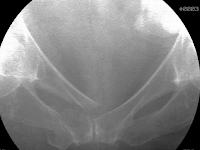
Her detailed neurological examination was otherwise normal. Pelvic plain radiographs and a computed tomography scan identified a right-sided displaced pubic ramus fracture and a displaced Y-shaped sacral fracture. On the anteroposterior image, she had a paradoxical inlet appearance of the sacrum.
The sagittal sacral CT scan images best demonstrated the displacement of the upper sacrum through the fracture’s transverse component.
She opted for surgical treatment of he unstable and displaced pelvic fractures. She underwent surgery the next day. After being anesthetized, she was positioned supine on a radiolucent table and elevated on a soft lumbosacral support. Manual compression of the pelvis under fluoroscopy identified the pubic ramus displacement due to instability.
Manipulative reduction using manual distraction at the iliac crests reduced the pubic fracture. Medullary fixation was performed percutaneously using a retrograde superior pubic ramus screw. The Y-shaped sacral fracture was stabilized percutaneously using two trans-iliac, trans-sacral iliosacral screws located safely within the upper sacral segment.
She had dramatic pain relief immediately after surgery. A licensed physical therapist monitored her rehabilitation for the next 3 months. For the initial six weeks after operation, light resistance strengthening exercises were selected along with right-sided protected weight bearing using a walker. Between weeks 7-12, progressive partial weight bearing and more vigorous strength training was performed. Four months after injury, she had returned to her normal activities without pain. Her plain pelvic radiographs demonstrated fracture union.
U, Y, and H-shaped sacral fractures are unusual and are often delayed diagnoses. The AP pelvic plain film may demonstrate a paradoxical inlet of the sacrum when the upper sacral component is displaced and kyphotic. Mid-sagittal sacral CT imaging better defines the fracture details. Percutaneous pelvic fixation is used when the fracture fragments and therefore osseus fixation pathways are adequately reduced/realigned.
Authored By: M.L. Chip Routt, Jr., M.D.


No comments:
Post a Comment