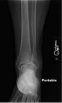
The Case for August 2012 is of a
35 year old male who sustained a fracture to his hip during a motorcycle
accident. He sustained a displaced
femoral neck fracture. He was seen by
the trauma team and was cleared for operative intervention for his fracture
once other injuries had been ruled out.


The fracture had both
medial and posterior superior comminution.
There was a retroversion deformity on the lateral view. This injury carries with it a significant
risk of developing avascular necrosis of the femoral head which can lead to
early arthritis and disability. Fracture
union can also be problematic as there are significant shear forces across the
fracture during healing.
Anatomic reduction in
a timely manner is key to maintaining blood flow to the femoral head and
allowing for the best chance of fracture healing. The patient was brought to the operating room
when cleared for an open reduction of his fracture. This was accomplished through a
Smith-Peterson anterior approach allowing for direct visualization of the
fracture. With this approach we were
able to clean the fracture, clamp it on the anterior tension side to restore
anteversion, and restore anatomic alignment under direct visualization. Also, the hip capsule was incised allowing
for decompression of the joint to hopefully improve blood flow to the head.


The
fracture reduction was held with a clamp and wire and then guide wires for 7.0
mm cannulated screws were placed through a separate small lateral incision. We avoided the posterior superior neck region
with our fixation so we would not further compromise blood flow to the femoral head. A screw was placed inferiorly
abutting the neck and then two further screws were placed more superiorly with
good spread. Partially threaded screws
were used to allow for fracture compression.


Final
intraoperative plain films showed an excellent reduction.


The patient was
allowed to perform toe-touch weightbearing for three months. He then progressed to full weight bearing as
tolerated. Serial radiographs were taken
throughout his follow-up at six weeks and three months showing no loss of
reduction.
The patient was seen
9 months following surgery. He walked
with a normal gait, had no hip pain, and had returned to all vocational and
avocational activities. Although the
patient is still at risk of developing avascular necrosis in the future,
radiographs showed a healed fracture with no evidence of avascular necrosis of the
femoral head at last follow-up.


A femoral neck fracture in a young patient is a serious injury as it can lead to pain and
arthritis secondary to avascular necrosis.
Anatomic reduction with secure fixation contributes to a successful
outcome.
Author by Daphne M. Beingessner, MD